A Semi-supervised Approach to Segment Retinal Blood Vessels in Color Fundus Photographs
Published in Conference on Artificial Intelligence in Medicine in Europe, 2019
Recommended citation: Sayed M.A., Saha S., Rahaman G.M.A., Ghosh T.K., Kanagasingam Y. (2019) A Semi-supervised Approach to Segment Retinal Blood Vessels in Color Fundus Photographs. In: Riaño D., Wilk S., ten Teije A. (eds) Artificial Intelligence in Medicine. AIME 2019. Lecture Notes in Computer Science, vol 11526. Springer, Cham https://link.springer.com/chapter/10.1007/978-3-030-21642-9_44
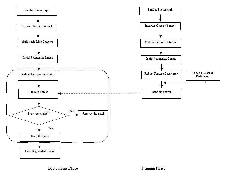
 Segmentation of retinal blood vessels is an important diagnostic procedure in ophthalmology. In this paper we propose an automated blood vessels segmentation method that combines both supervised and un-supervised approaches. A novel descriptor named Local Haar Pattern (LHP) is proposed to describe retinal pixel of interest. The performance of the proposed method has been evaluated on three publicly available DRIVE, STARE and CHASE_DB1 datasets. The proposed method achieves an overall segmentation accuracy of 96%, 96% and 95% respectively on DRIVE, STARE, and CHASE DB1 datasets, which are better than the state-of-the-art methods.
Download paper here

